Zoology Anatomy Models
Showing 1–24 of 26 results
- Zoology Anatomy Models
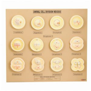
Animal Cell Division Meiosis Zoology Model
Animal Cell Division Meiosis Zoology Model – Set of 12 models, showing detailed stages in Meiosis cell division, beautifully coloured to bring out the detailed structure as seen under the microscope, mounted on base with key card.
SKU: n/a - Zoology Anatomy Models
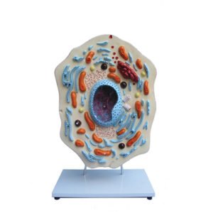
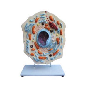
Animal Cell Zoology Model
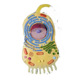
This model, enlarged about 20,000 times. Animal Cell Model, provides clear depiction of the delicate and complex structure of the animal cell. Shows the nucleus , endoplasmatic reticulum, mitochondria, ribosomes, polysomes and Golgi apparatus, centrioles, lysosomes and fat vacuoles.
SKU: n/a - Zoology Anatomy Models
Cat Dissection Model
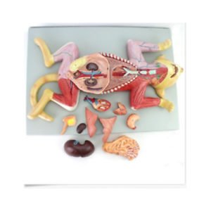
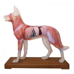
This life-size dissection model features over 100 individual anatomical details. An extremely realistic and precise model, it is crafted with hand-painted detail. Featured structures include a cross-sectioned kidney showing the cortex and medulla, major arteries and veins, muscle groups of the fore and hind limbs, and the open uterus exposing a developing fetus. This model also has an open mouth cavity detailing the teeth and nasopharynx, and includes a key identifying 136 structures.
SKU: n/a - Zoology Anatomy Models
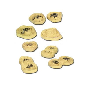
Frog Life Cycle Specimens
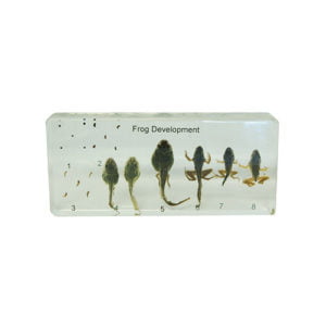
Frog Life Cycle Specimens – Frog Development specimens in presentation box showing different stages of the frog life cycle. Information card included. Specimens include egg, tadpole 6 weeks, tadpole with legs 9 weeks, tadpole with 4 limbs 10 weeks, tadpole with short tail 11 weeks, froglet and adult frog. These durable and clear specimens in lightweight acrylic provide students with a fascinating insight into animal life.
SKU: n/a - Zoology Anatomy Models
Paramecium Enlarged Zoology Model
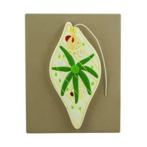
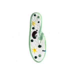
Paramecium, enlarged approx. 1600 times, showing the cell inventory of a protozoa: macro and micronucleus, contractile vacuoles, cytostome with membranellae, myonemes and food vacuoles and the formation of the endo and ectoplasm and the network of neuronemes, it also shows the structure of the pellicle of the ectoplasm, and the position and order of the trichocysts and a range of cilia in typical order, mounted on base with key card.
SKU: n/a

